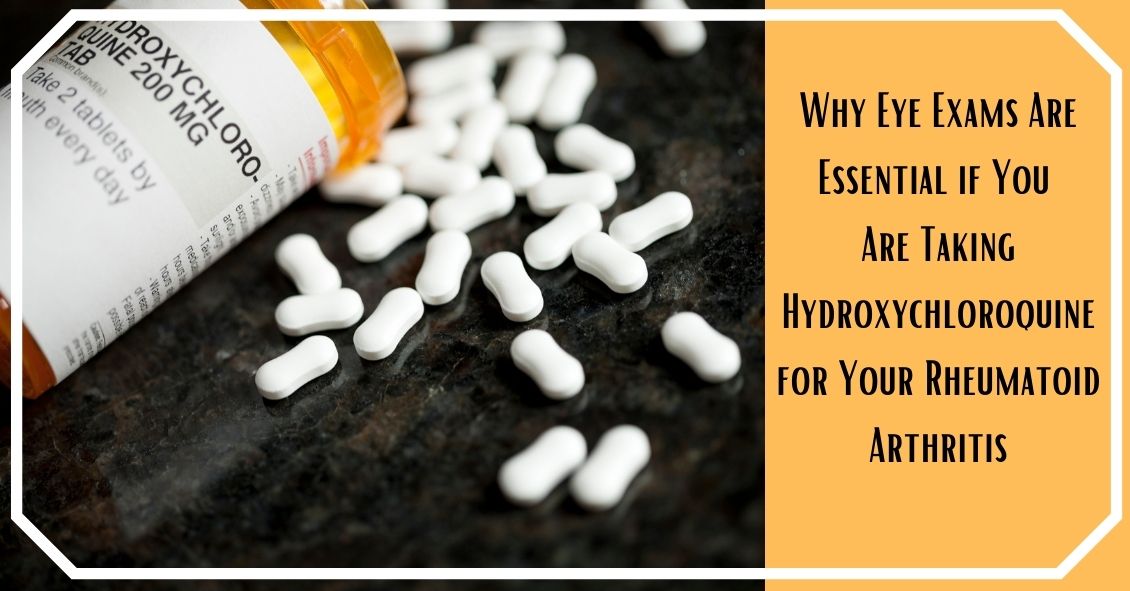
Hydroxychloroquine (Plaquenil) was originally used to treat malaria and is now used mostly to treat rheumatological and dermatological diseases. Its most frequent use now is for rheumatoid arthritis (RA) and Lupus and is often very effective in mitigating the joint and arthritic symptoms these diseases can cause.
One of the most significant side effects of the drug is its possibility of causing eye problems resulting in blurred or decreased vision. The most common issue is damage to the retina. It can impair your color vision or damage the retinal cells, particularly in the area right around the central vision.
In your retina, the area that you use to look straight at an object is called the fovea. The fovea is the area that provides you with the most definition when looking at an object. The area just around the fovea is called the macula and it has the ability to see objects with slightly less definition than the fovea but significantly better than the rest of your retina, which accounts for your peripheral vision. The most common place for Hydroxychloroquine to cause a problem is in a ring of the macula surrounding the fovea.
The reason it is important to detect any of these changes as early as possible is because in many instances the changes are not reversible even if you come off the medication.
The risk of this happening is highly correlated with the cumulative dose of the drug you have received. So, the higher the dose and the longer you have been on it the higher your risk.
The current recommendation is a daily dose that does not exceed 6.5 mg/kg/day (that is milligrams per kilograms per day). There are approximately 2.2 pounds. in a kilogram. The pills come in 200 mg tablets. Most people who are on this drug are on either 200 mg once a day or 200 mg twice a day. The safety break point comes at around 135 pounds. People weighing more than that will stay within the safety guidelines (not more than 6.5mg/kg/day) at 400mg per day, but people under 135 pounds should probably only be taking 200 mg per day.
Other risk factors for Hydroxychloroquine retinal toxicity include kidney or liver disease and obesity. Obesity is a risk factor because the drug does not penetrate fat tissue so there is more of the drug in your lean body mass (including your retina and its supporting cells called the retinal pigment epithelium). What that means in real terms is that if you take two people who each weigh 140 pounds and put them both on 400 mg a day and one person is 4-foot 11 and the other is 5-foot 9, the 4-foot 11 inch person is at greater risk for side effects because the shorter person has more of their body weight in fat tissue. Since the hydroxychloroquine can’t penetrate the fat tissue that means there is a higher concentration of it in sensitive tissues like the retina. People with kidney and liver problems have a tougher time eliminating the drug from their system so they are at higher risk because the body is going to retain more of the drug for a longer period of time.
The recommendation is to have a baseline eye exam with dilation and a visual field test before or soon after starting the drug. A repeat of that exam should occur every year if there is no evidence of toxicity.
The actual incidence of retinal toxicity from hydroxychloroquine is difficult to pin down because there is usually a long time between being started on the drug and the start of any identifiable retinal toxicity. The overall rate of probable retinal toxicity is in the range of 1 of every 200 people treated. The rate is much lower than that in the first 7 years of treatment but gets to about 5 times higher after 7 years of treatment. Some of that data is old now and there is much greater awareness currently about keeping people below that 6.5 mg/kg/day dosage level.
I have been in practice for over 25 years and have seen “probable” retinal toxicity from hydroxychloroquine a total of 5 times and only once in the last 10 years when people have been more careful about keeping the dosage in the right range.
The drug can be very effective in its treatment of RA and Lupus and the likelihood of serious vision problems is small and can potentially be avoided with the correct dosing and monitoring of the eyes. Other drugs in the treatment for RA or Lupus may have more frequent or serious side effects then Hydroxychloroquine so it would be wise to consider it a viable treatment option and not easily dismiss it because of the risk of what amounts to a fairly infrequent eye issue.
Article contributed by Dr. Brian Wnorowski, M.D.
Read more: Rheumatoid Arthritis, Hydroxychloroquine, and Your Eyes
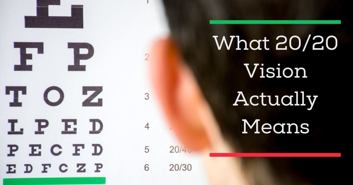
One of the most commonly asked questions in an eye exam comes right after the refraction, or glasses prescription check: “What is my vision?”
Almost invariably, people know the term “20/20”. In fact, it’s a measure of pride for many people. “My doctor says I have 20/20 vision.” Or, on the other side of that same coin, having vision that is less than 20/20, say 20/400, can be a cause of great concern and anxiety. In this discussion I will describe what these terms actually mean.
To lay the foundation, let’s discuss some common terms. Visual acuity (VA) is clarity or sharpness of vision. Vision can be measured both corrected (with glasses or contact lenses) and uncorrected (without glasses or contact lenses) during the course of an eye exam. The result of an eye exam boils down to two different but related sets of numbers: your VA and your actual glasses prescription.
The notation that doctors use to measure VA is based off of a 20-foot distance. This is where the first 20 in 20/20 comes from. In Europe, since they use the metric system, it is based on meters. The 20/20 equivalent is 6/6 because they use a 6-meter test distance. The second number is the smallest line of letters that a patient can read. In other words, 20/20 vision means that at a 20-foot test distance, the person can read the 20/20 line of letters.
The technical definition of 20/20 is full of scientific jargon - concepts such as minutes of arc, subtended angles, and optotype size. If you’d like to read more of the technical details there is a well-written article with illustrations by Dr. John Ellman, you can find here. For the purposes of our discussion here I’ll try to explain it in less technical terms.
“Normal” vision is somewhat arbitrarily set as 20/20 (some people can see better than that). Let’s say you have two people: Person A with 20/20 vision and Person B with 20/40 vision. The smallest line of letters that person B can see at 20 feet is the 20/40 line. Person A, with “normal” 20/20 vision, could stand 40 feet away from that same line and see it. There is somewhat of a linear relationship in that the 20/40 letters are twice the size of the 20/20 letters and someone with normal vision could see a 20/40 letter at twice the distance as the person with 20/40 vision.
So how does this translate to a glasses prescription?
Eye doctors can often estimate what your uncorrected VA will be based on your glasses prescription. This works mainly for near-sightedness. Essentially, every quarter step of increasing glasses prescription (i.e. -1.25 as compared to -1.50) means a person can see one less line on a VA chart.
A prescription of - 1.25 works out to roughly 20/50 vision, -1.50 to 20/60 and so on. Anybody with an anatomically sound eyeball, meaning the absence of any kind of disease process, should generally be correctable to 20/20 with glasses or contact lenses. It is important to note, however, that rarely a person’s best corrected VA may be less than 20/20 with no noticeable signs of disease.
Far-sightedness is more difficult to estimate because it is affected by a number of other factors, including one’s age and focusing ability. But that’s a topic for another article.
So there you have it! Hopefully this has shed some light on what these measurements that we take actually mean, and it has allowed you to understand your eye health a little bit better.
Article contributed by Dr. Jonathan Gerard

Here are 11 bad contact lens habits we eye doctors often see--
#1 Sleeping in your contacts.
This is the No. 1 risk factor for corneal ulcers, which can lead to severe vision loss and the need for a corneal transplant. Your cornea needs oxygen from the atmosphere because it has no blood vessels. The cornea is already somewhat deprived of oxygen when you have your eyes closed all night, and adding a contact on top of that stresses the cornea out from lack of oxygen. You don’t need to see when you are sleeping. Take your contacts out!!! I promise your dreams will still look the same.
#2 Swimming in your contacts.
Salt, fresh, or pool water all have their individual issues with either bacteria or chemicals that can leach into your contacts. If you absolutely need to wear them to be safe in the water, then take them out as soon as you are done and clean and disinfect them.
#3 Using tap water to clean contacts.
Tap water is not sterile. See No. 2.
#4 Using your contacts past their replacement schedule.
The three main schedules now are daily, two weeks, and monthly. Dailies are just that – use them one time and then throw them away. They are not designed to be removed and re-used. Two-week contacts are designed to be thrown away after two weeks because they get protein buildup on them that doesn’t come off with regular cleaning. Monthly replacement contacts need to have both daily cleaning and weekly enzymatic cleaning to take the protein buildup off. Using your lenses outside of these schedules and maintenance increases the risk of infection and irritation.
#5 Getting contacts from an unlicensed source.
Costume shops and novelty stores sometimes illegally sell lenses. If you didn’t get the fit of the lenses checked by an eye doctor, they could cause serious damage if they don’t fit correctly.
#6 Wearing contacts past their expiration date.
You can’t be sure of the sterility of the contact past its expiration date. As cheap as contacts are now, don’t take the risk with an expired one.
#7 Topping off your contact lens case solution instead of changing it.
This is a really bad idea. Old disinfecting solution no longer kills the bacteria and can lead to resistant bacteria growing in your case and on your lenses that even fresh disinfecting solution may not kill. Throw out the solution in the case EVERY DAY!
#8 Not properly washing your hands before inserting or removing contacts.
It should be self-evident why this is a problem.
#9 Not rubbing your contact lens when cleaning even with a “no rub” solution.
Rubbing the lens helps get the bacteria off. Is the three seconds it takes to rub the lens really that hard? “No rub” should never have made it to market.
#10 Sticking your contacts in your mouth to wet them.
Yes, people actually do this. Do you know the number of bacteria that reside in the human mouth? Don’t do it.
#11 Not having a backup pair of glasses.
This is one of my biggest pet peeves with contact lens wearers. In my 25 years of being an eye doctor, the people who consistently get in the biggest trouble with their contacts are the ones who sleep in them and don’t have a backup pair of glasses. So when an eye is red and irritated they keep sticking that contact lens in because it is the only way they can see. BAD IDEA. If your eye is red and irritated don’t stick the contact back in; it’s the worst thing you can do!
Article contributed by Dr. Brian Wnorowski, M.D.
The content of this blog cannot be reproduced or duplicated without the express written consent of Eye IQ

The jury is still out on that question. There is some supportive experimental data in animal models but no well-done human studies that show significant benefit.
What you shouldn’t do is pass up taking the AREDS 2 nutritional supplement formula, which is clinically proven to reduce the risk of severe visual loss that can happen with macular degeneration. Almost all the data supporting the POSSIBLE benefits of bilberry in visual conditions is related to NON-HUMANS. Stick with the AREDS 2 formula that has excellent clinical evidence.
So, what is bilberry and why do some people use it?
Bilberry (Vaccinium myrtillus), a low-growing shrub that produces a blue-colored berry, is native to Northern Europe and grows in North America and Asia. It is naturally rich in anthocyanins, which have anti-oxidant properties.
It is said that during World War II, British pilots in the Royal Air Force ate bilberry jam, hoping to improve their night vision. No one is exactly sure where the impetus to do this came from, but it is believed that this story is what lead to some widespread claims that bilberry was good for your eyes.
A study by JH Kramer, Anthocyanosides of Vaccinium myrtillus (Bilberry) for Night Vision - A Systematic Review of Placebo-Controlled Trials, reviewed most of the literature pertaining to the claim that bilberry improves night vision. He found that the four trials, which were all rigorous randomized controlled trials (RCTs), showed no correlation with bilberry extract and improved night vision. A fifth RCT and seven non-randomized controlled trials reported positive effects on outcome measures relevant to night vision, but these studies had less-rigorous methodology.
Healthy subjects with normal or above-average eyesight were tested in 11 of the 12 trials. The hypothesis that V. myrtillus improves normal night vision is not supported by evidence from rigorous clinical studies. There is a complete absence of rigorous research into the effects of the extract on subjects suffering impaired night vision due to pathological eye conditions.
Even though there is no solid evidence in human studies that bilberry produces any positive visual effects on night vision there is some experimental evidence that implies it might be useful in some ocular conditions whose mode of action is oxidative stress. There are recent epidemiologic, molecular and genetic studies that show a major role of oxidative stress in age-related macular degeneration.
There have been some studies showing oxidative protective effects of bilberry in non-human models.
In Protective Effects of Bilberry and Lingonberry Extracts Against Blue-light Emitting Diode Light-induced Retinal Photoreceptor Cell Damage in Vitro, Ogawa et al showed that cultured mouse cells that had bilberry extract added before subjection to high-energy short-wavelength light survived better than those that hadn't received the extract.
In Retinoprotective Effects of Bilberry Anthocyanins via Antioxidant, Anti-Inflammatory, and Anti-Apoptotic Mechanisms in a Visible Light-Induced Retinal Degeneration Model in Pigmented Rabbits, Wang et al found similarly improved survival of pigmented rabbit retinal cells when exposed to bilberry abstract prior to high-intensity light.
But bilberry is not without potential side effects.
Bilberry possesses anti-platelet activity and it might interact with NSAIDs, particularly aspirin. Excessive drinking of bilberry juice might cause diarrhea. One study of 2,295 people given bilberry extract found a 4% incidence of side effects or adverse events. Further, bilberry side effects may include mild digestive distress, skin rashes and drowsiness. Chronic uses of the bilberry leaf may lead to serious side effects. High doses of bilberry leaf can be poisonous.
Bilberry has not been evaluated by the Food and Drug Administration for safety, effectiveness, or purity.
Article contributed by Dr. Brian Wnorowski, M.D.

You’ve been diagnosed with a cataract and you’ve been told you should have cataract surgery. The surgeon is also telling you that you should consider paying extra out-of-pocket for it.
Where did this come from? Why should you have to pay out-of-pocket for cataract surgery? Shouldn’t your health insurance just cover it?
In trying to answer these questions, you will first need a little history of both cataract and refractive surgery, which corrects errors of refraction such as nearsightedness, farsightedness, and astigmatism.
Radial keratotomy (RK) was the first widely used refractive surgery for nearsightedness. It was invented in 1974 by Russian ophthalmologist Svyatoslav Fyodorov, and it was the primary refractive procedure done until the mid-1990s. Then it was surpassed by the laser procedure called PRK and then, eventually, LASIK; they are still the predominately pure refractive surgeries done today.
Cataract surgery has its origins all the way back to at least 800 BC in a procedure called couching. In this procedure, the cataract was pushed into the back of the eye with a sharp instrument so the person could look around the cataract. Medically that is all that was done with cataracts until around 1784 when a cataract was actually removed from the eye.
The next big advance was implants to replace the removed cataract. The invention of implants was spurred by Harold Ridley, who recognized that injured Royal Air Force pilots could retain shards of their canopy made out of a substance called PMMA in their eye without the body rejecting it. Implants became commonplace after the FDA approved them in 1981. The implants have improved over the years and most implants today are foldable, enabling them to fit through tiny incisions of around 3 millimeters.
Medicare and most other insurances cover the cost of MEDICALLY NECESSARY cataract surgery. This means they will cover the surgery when someone has symptoms of visual trouble that is interfering with their normal daily activities AND the cataract is the cause of those visual disturbances. There is no reason to remove a cataract just because it is there. It needs to be causing a problem to make it medically necessary to remove it.
Medicare and most other insurance do not cover refractive surgery (LASIK, PRK, etc.). The general perception of refractive surgery by the insurance industry is that it is not MEDICALLY NECESSARY. You can correct the refractive errors in almost all cases by non-surgical means, such as glasses an/ or contact lenses.
Today there are methods of doing additional procedures, or using special implants, at the time of cataract surgery to correct more than just the cataract alone. This is where the two types of surgeries, refractive and cataract, have merged into a single operation that tries to take care of both problems.
The merging of cataract and refractive surgeries is why there are now options to not only get your cataract removed, but also to correct your astigmatism (irregular shape to cornea) and/or presbyopia (the inability to see well up close that hits nearly everyone in their 40’s).
This is where the "paying for cataract surgery" comes in. Surgery to correct astigmatism and presbyopia are not considered MEDICALLY NECESSARY because they can be corrected with eyeglasses or contacts.
Your cataract, once it hits a certain point, cannot be corrected with glasses or contacts and therefore it is MEDICALLY NECESSARY and your insurance will pay for that component of your surgery. What it won’t pay for is any additional amount that is charged to correct your astigmatism or presbyopia.
If you want to address your astigmatism and/or presbyopia at the time of cataract surgery in order to be less dependent on wearing glasses after surgery, then paying for those components is going to be an out-of-pocket payment for you.
Article contributed by Dr. Brian Wnorowski, M.D.
This blog provides general information and discussion about eye health and related subjects. The words and other content provided in this blog, and in any linked materials, are not intended and should not be construed as medical advice. If the reader or any other person has a medical concern, he or she should consult with an appropriately licensed physician. The content of this blog cannot be reproduced or duplicated without the express written consent of Eye IQ.
Read more: I Should Pay Out-of-Pocket for Cataract Surgery Now?
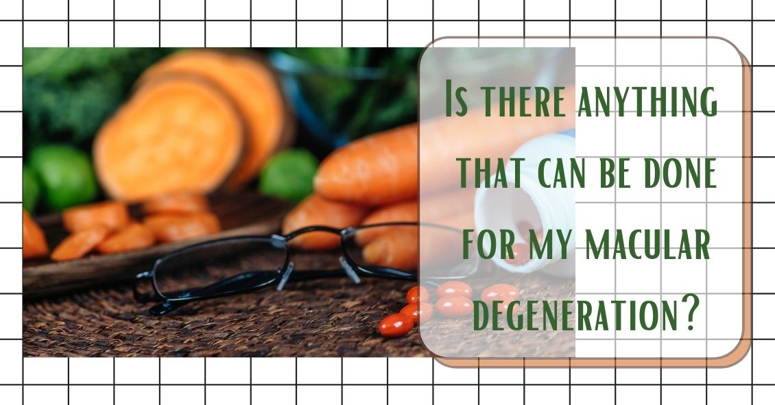
Here are some treatment options for Dry and Wet Age Related Macular Degeneration.
Nutritional supplements and Dry Age Related Macular Degeneration (AMD)
The Age-Related Eye Disease Study 2 (AREDS2) showed that people at high risk of developing advanced stages of AMD benefited from taking dietary supplements. Supplements lowered the risk of macular degeneration progression by 25 percent. These supplements did not benefit people with early AMD or people without AMD.
Following is the supplementation:
- Vitamin C - 500 mg
- Vitamin E - 400 IU
- Lutein – 10 mg
- Zeaxanthin – 2 mg
- Zinc Oxide – 80 mg
- Copper – 2 mg (to prevent copper deficiency that may be associated with taking high amount of zinc)
Another study showed a benefit in eating dark leafy greens and yellow, orange and other fruits and vegetables. These vitamins and minerals listed above are recommended in addition to a healthy, balanced diet.
It is important to remember that vitamin supplements are not a cure for AMD, nor will they restore vision. However, these supplements may help some people maintain their vision or slow the progression of the disease.
Wet AMD treatments
The most common treatment for wet AMD is an eye injection of anti-vascular endothelial growth factor (anti-VEGF). This treatment blocks the growth of abnormal blood vessels, slows their leakage of fluid, may help slow vision loss, and in some cases can improve vision. There are multiple anti-VEGF drugs available: Avastin, Lucentis, and Eylea.
You may need monthly injections for a prolonged period of time for treatment of wet AMD.
Laser Treatment for Wet AMD
Some cases of wet AMD may benefit from thermal laser. This laser destroys the abnormal blood vessels in the eye to prevent leakage and bleeding in the retina. A scar forms where the laser is applied and may cause a blind spot that might be noticeable in your field of vision.
Photodynamic Therapy or PDT
Some patients with wet AMD might benefit from photodynamic therapy (PDT). A medication called Visudyne is injected into your arm and the drug is activated as it passes through the retina by shining a low-energy laser beam into your eye. Once the drug is activated by the light it produces a chemical reaction that destroys abnormal blood vessels in the retina. Sometimes a combination of laser treatments and injections of anti-VEGF mediations are employed to treat wet AMD.
Article contributed by Jane Pan M.D.
The content of this blog cannot be reproduced or duplicated without the express written consent of Eye IQ
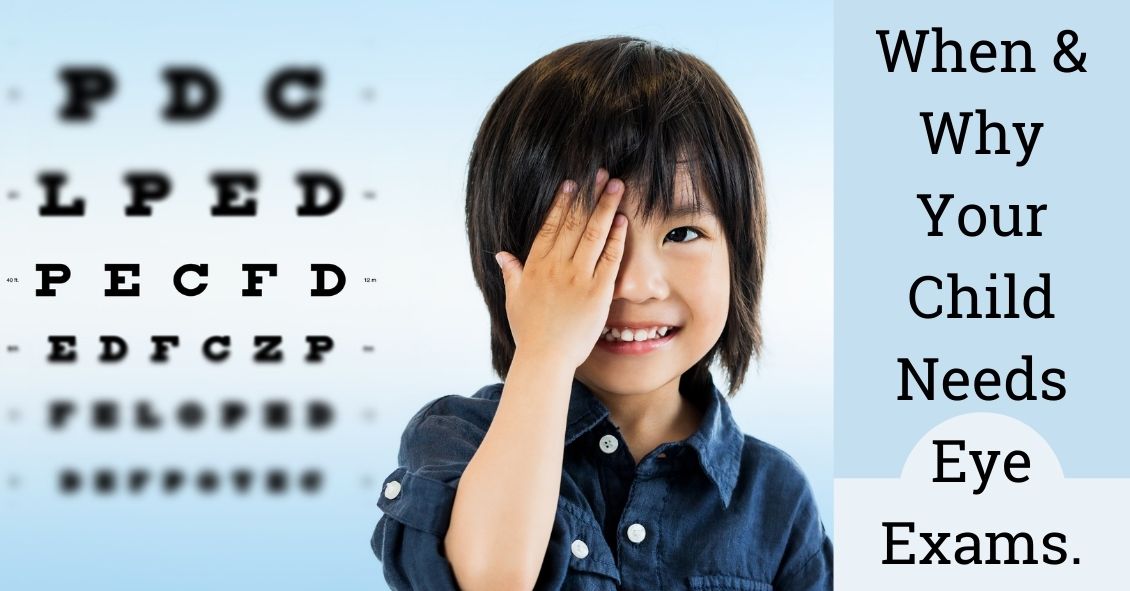
Just like adults, children need to have their eyes examined. This need begins at birth and continues through adulthood.
Following are common recommendations for when a child needs to be screened, and what is looked for at each stage.
A child’s first eye exam should be done either right at or shortly after birth. This is especially true for children who were born prematurely and have a very low birth weight and may need to be given oxygen. This is mainly done to screen for a disease of the retina called retinopathy of prematurity (ROP), in which the retina does not develop properly as a result of the child receiving high levels of oxygen. Although rarer today due to the levels being monitored more closely, it is still a concern for premature babies.
The next time an eye exam is in order is around 6 months. At this stage, your pediatric eye doctor will check your child’s basic visual abilities by making them look at lights, respond to colors, and be able to follow a moving object.
Your child’s ocular alignment will also be measured to ensure that he or she does not have strabismus, a constant inward or outward turning of one or both eyes. Parents are encouraged to look for these symptoms at home because swift intervention with surgery to align the eyes at this stage is crucial for their ocular and visual development.
It is also imperative for parents and medical professionals to be on the lookout for retinoblastoma, a rare cancer of the eye that more commonly affects young children than adults. At home, this might show up in a photo taken with a flash, where the reflection in the pupil is white rather than red. Other symptoms can include eye pain, eyes not moving in the same direction, pupils always being wide open, and irises of different colors. While these symptoms can be caused by other things, having a doctor check them immediately is important because early treatment can save your child’s sight, but advanced cases can lead to vision loss and possibly death if the cancer spreads.
After the 6-month exam, I usually recommend another exam around age 5, then yearly afterward. There are several reasons for this gap. First, any parent with a 2- to 4-year-old knows that it’s difficult for them to sit still for anything, let alone an eye exam. Trying to examine this young of a patient can be frustrating for the doctor, the parent, and the child. Nobody wins. By age 5, children are typically able to respond to questions and can (usually) concentrate on the task at hand. If necessary at this stage, their eyes will be measured for a prescription for glasses and checked for amblyopia, commonly known as a “lazy eye”. Detected early enough, amblyopia can be treated properly under close observation by the eye doctor.
The recommendations listed above are solely one doctor’s opinion of when children should have eye exams. The various medical bodies in pediatrics, ophthalmology, and optometry have different guidelines regarding exam frequency, but agree that while it is not essential that a healthy child’s eyes be examined every year, those with a personal or family history of inheritable eye disease should be followed more closely.
Article contributed by Dr. Jonathan Gerard
NOTE: Many eye doctors commonly like to have another exam around age 3, in order to make sure a pre-schooler's vision is developing correctly. Please go by what your trusted eye doctor advises.
This blog provides general information and discussion about eye health and related subjects. The words and other content provided on this blog, and in any linked materials, are not intended and should not be construed as medical advice. If the reader or any other person has a medical concern, he or she should consult with an appropriately licensed physician. The content of this blog cannot be reproduced or duplicated without the express written consent of Eye IQ.
The Background
Over the last several years, research has indicated a strong correlation between the presence of Obstructive Sleep Apnea (OSA) and glaucoma. Information from some of these pivotal studies is presented below.
Did you know
- Glaucoma affects over 60 million people worldwide and almost 3 million people in the U.S.
- There are many people who have glaucoma but have not yet had it diagnosed.
- Glaucoma is a leading cause of blindness in the United States.
- If glaucoma is not detected and goes untreated, it will result in peripheral vision loss and eventual, irreversible blindness.
- Sleep apnea is a condition that obstructs breathing during sleep.
- It affects 100 million people around the globe and around 25 million people in the U.S.
- A blocked airway can cause loud snoring, gasping or choking because breathing stops for up to two minutes.
- Poor sleep due to sleep apnea results in morning headaches and chronic daytime sleepiness.
The Studies
In January 2016, a meta-analysis by Liu et. al., reviewed studies that collectively encompassed 2,288,701 individuals over six studies. Review of the data showed that if an individual has OSA there is an increased risk of glaucoma that ranged anywhere from 21% to 450% depending on the study.
Later in 2016, a study by Shinmei et al. measured the intraocular pressure in subjects with OSA while they slept and had episodes of apnea. Somewhat surprisingly they found that when the subjects were demonstrating apnea during sleep, their eye pressures were actually lower during those events than when the events were not happening.
This does not mean there is no correlation between sleep apnea and glaucoma - it just means that an increase in intraocular pressure is not the causal reason for this link. It is much more likely that the correlation is caused by a decrease in the oxygenation level (which happens when you stop breathing) in and around the optic nerve.
In September of 2016, Chaitanya et al. produced an exhaustive review of all the studies done to date regarding a connection between obstructive sleep apnea and glaucoma and came to a similar conclusion. The risk for glaucoma in someone with sleep apnea could be as high as 10 times normal. They also concluded that the mechanism of that increased risk is most likely hypoxia – or oxygen deficiency - to the optic nerve.
The Conclusion
There seems to be a definite correlation of having obstructive sleep apnea and a significantly increased risk of getting glaucoma. That risk could be as high as 10 times the normal rate.
It's highly recommended that if you have been diagnosed with obstructive sleep apnea that you have have a comprehensive eye exam in order to detect your potential risk for glaucoma.
Article contributed by Dr. Brian Wnorowski, M.D.
This blog provides general information and discussion about eye health and related subjects. The words and other content provided on this blog, and in any linked materials, are not intended and should not be construed as medical advice. If the reader or any other person has a medical concern, he or she should consult with an appropriately licensed physician. The content of this blog cannot be reproduced or duplicated without the express written consent of Eye IQ.
Do you have family members with eye-related conditions?
The two main eye diseases in adults that have a genetic link are glaucoma and age-related macular degeneration (AMD).
Glaucoma is a deterioration of the optic nerve caused by pressure in the eye or poor blood flow to the optic nerve. It has no symptoms at its onset. In most cases if you wait until you begin to realize there is something wrong with your vision to get glaucoma diagnosed, upwards of 70% of your optic nerve will have already been destroyed. Once the nerve is destroyed there is no way of reversing that today and treatment is focused on trying to preserve whatever nerve tissue is left.
Your chances of getting glaucoma are four to 10 times higher if you have a close relative with glaucoma. Getting your eyes examined regularly is always important but even more so if there is a family history of glaucoma.
Macular degeneration is the leading cause of blindness in most of the developed world. It too can cause serious vision loss if you wait until you have significant symptoms before a diagnosis. There are now some preventative treatments for AMD--the earlier it is detected the better off you will be.
Having a close family member with AMD may increase your chances of having the disease as much as 50 percent, making timely diagnosis and treatment imperative.
Other eye diseases that run in families include strabismus (crossed eyes), myopia (nearsightedness), hyperopia (farsightedness), and astigmatism.
All of these family connections are important to know so that you and your eye doctor can together take the best possible care of your eyes. Before your next eye exam ask your relatives if they have any history of eye disease. It might not make for the lightest of conversation at your next family gathering but it could help save your vision.
Article contributed by Dr. Brian Wnorowski, M.D.
The content of this blog cannot be reproduced or duplicated without the express written consent of Eye IQ

Itching, burning, watering, red, irritated, tired eyes... what is a person to do? The symptoms aforementioned are classic sign of Dry Eye Syndrome (DES), which affects millions of adults and children. With increased screen time in all age groups, the symptoms are rising.
What causes this? One reason is that when we stare at a computer screen or phone, our blink reflex slows way down. A normal eye blinks 17,000 times per day. When our eye functions normally, the body usually produces enough tears to be symptom free, however, if you live in a geographical area that is dry, or has a high allergy rate, your symptoms could be worse.
Dry eye syndrome can be brought on by many factors: aging, geographical location, lid hygiene, contact lens wear, medications, and dehydration. The lacrimal gland in the eye that produces tears, in a person over forty years old, starts slowly losing function. Females with hormonal changes have a higher incidence of DES (dry eye syndrome). Dry, arid climates or areas with high allergy causes lend to higher incidences of DES as well.
Blepharities, a condition of the eyelids, can cause a dandruff-like situation for the eye, exacerbating a dry eye condition. Contact lenses can add to DES, so make sure you are in high oxygen contact lens material if you suffer from DES. Certain medications such as antihistamines, cholesterol and blood pressure meds, hormonal and birth control medications, can also cause symptoms of a dry eye. Check with your pharmacist if you are not sure.
And finally, overall dehydration can cause DES. Some studies show we need 1/2 our body weight in ounces of water per day. For example, if you weigh 150 lbs, you need approximately 75 ounces of water per day to be fully hydrated. If you are not at that level, it could affect your eyes.
Treatment for DES is varied, but the main treatment is a tear supplement to replace the evaporated tears. These come in the form of topical ophthalmic artificial tears. Oral agents that can help are Omega 3 supplements such as fish oil or flax seed oil pills. They supplement the function of meibomian glands located at the lid margin. Ophthalmic gels used at night, as well as humidifiers, can help keep your eyes moisturized. Simply blinking hard more often can cause the lacrimal gland to produce more tears automatically.
For stubborn dry eyes, retaining tears on the eye can be aided by punctal plugs. They act like a stopper for a sink, and they are painless and can be inserted by your eye care practitioner in the office. Moisture chamber goggles can also be used in severe cases to hydrate the eyes with their body’s own natural humidity. This may sound far out but it gets the job done.
Being aware of the symptoms and treatments for dry eye syndrome can prevent frustration and allow your eyes to work more smoothly and efficiently in your daily routine. If your eyes feel dry as the Sahara or they water too much, know that help is on the way through proven techniques and products. You do not need to suffer needlessly in the case of Dry Eye Syndrome anymore. Make an appointment to talk with your eye doctor about the best treatments for you!
This blog provides general information and discussion about eye health and related subjects. The words and other content provided in this blog, and in any linked materials, are not intended and should not be construed as medical advice. If the reader or any other person has a medical concern, he or she should consult with an appropriately licensed physician. The content of this blog cannot be reproduced or duplicated without the express written consent of Eye IQ.
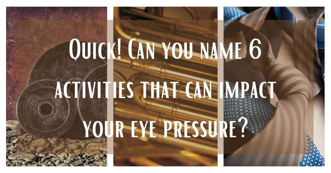
There have been studies undertaken over the past several years to try and understand if there are any of our day-to-day activities that either help or hurt the management of glaucoma.
Most of the studies demonstrated very little impact on the course of glaucoma. Here are some of the things researches have looked at.
Aerobic exercise: This means doing something at least four times per week for more than 20 minutes at a time that raises your pulse rate to a level that makes your heart work harder. Going from a sedentary lifestyle to active one with aerobic exercise resulted in a very slight decrease in baseline eye pressure.
Yoga: A study conducted at the Mount Sinai Health System (https://journals.plos.org/plosone/article?id=10.1371/journal.pone.0144505) showed a significant increase in eye pressure with any head-down positioning. People with glaucoma would be wise to avoid any exercise that involves a position where your head is lower than your heart.
Weight lifting: Holding your breath while exerting yourself (called the Valsalva maneuver), is also a time when your eye pressure can go sky high. So if you lift weights for exercise, which is generally a good idea to maintain bone density, make it low weights with more repetitions of lifting, rather than heavy weights that make you grunt.
Wind instruments: A similar breath-holding problem applies to those playing the larger wind musical instruments like the French horn. One study suggested that there was a greater chance of glaucoma in symphonic wind players. If you play a brass instrument, it makes sense to have frequent checks of pressure, optic nerve head, and visual field.
Marijuana: Smoking marijuana can lower eye pressure. However, due to its short duration of action (3-4 hours), side effects, and lack of evidence that it alters the course of glaucoma, it is not recommended for glaucoma treatment.
Wearing tight neckties: This creates a very short-duration increase in eye pressure but doesn’t seem to have any long-term effects.
Visual Field
The visual field test is designed to see how well you see outside of the center part of your vision (peripheral vision).
When we test your vision on the basic eye chart it is only testing how well you see right in the center and gives us no idea if you can see out away from the center. Your peripheral vision is very important because it gives you the ability to move around your environment without running into things.
There are several diseases that can severely impact your peripheral vision while leaving central vision unaffected. Some people can have perfectly normal 20/20 central visual acuity and have almost complete loss of their peripheral vision.
The main culprits that can have a big impact on your peripheral vision long before your central vision are glaucoma, some retinal diseases such as retinal detachments or retinitis pigmentosa, and some neurological problems like brain tumors, strokes, pseudotumor cerebri and multiple sclerosis.
Most visual field tests are now done on an automated machine that flashes lights in your peripheral vision while you stare straight ahead. The lights continue to get dimmer until you can no longer detect that they are there. The machine is trying to find the dimmest light you can see at each point in your peripheral vision that it is testing for.
Many patients get anxious when they take this test because everyone wants to do well on it. That sometimes results in people not staring straight ahead but trying to look around to find the lights in an effort to do better.
That just makes the test come out worse. The machine also makes some noise as it changes location of the test light. Some people start pressing the buzzer whenever they hear a noise. They think there must be a light they missed but the machine, several times during the test, makes noise and then doesn’t put a light on to specifically see if you are trying to cheat by hitting the buzzer on the noise rather that seeing the light. Don’t do those things - you are only cheating yourself and making it more difficult to figure out your problem.
Ocular Coherence Tomography (OCT)
The OCT really took hold in eye doctors' offices at the beginning of this century. It was the first time we were able to see anatomy and pathology inside the eye on a microscopic level without the use of any radiation.
It has been a great addition to our examination techniques and allowed us to see many causes of vision loss at a level of detail we could never have before.
The two biggest uses for OCT in optical health are diagnosing diseases of the retina, particularly the area of central vision called the macula, and for diseases of the optic nerve, the most common of which is glaucoma.
For retinal disease it has been extremely helpful in macular problems such as macular degeneration, the leading cause of blindness in the U.S., diabetic retinopathy, retinal vascular occlusions and retinal swelling from inflammation.
The OCT allows us to see the individual cellular levels of the retina and helps in diagnosing the exact level where the pathology is occurring. If you look into the eye at the retina and see some bleeding in the macula it is difficult to judge where that blood exists. Is it superficial in the retina and coming from the retinal circulation or is it deep in and coming from the choroidal circulation under the retina?
The difference between those two locations can have a significant impact on what disease is causing the problem and what the proper treatment is. The OCT is also very helpful in following the effect of treatment. If you are treating a bleeding or swelling problem in the retina, the OCT can track the degree of improvement with a level of detail that could never be matched by the human eye.
For glaucoma and other problems with the optic nerve, the OCT can very precisely measure the thickness of the nerve tissue as it passes through the optic nerve. The hallmark of glaucoma is progressive loss of nerve fibers in the optic nerve. Being able to measure the nerve thickness down to the micron level is very helpful in both diagnosing and watching for progression of any optic nerve disease.
Fundus Photography
A picture is worth 1,000 words...
Fundus photography is just that, a regular picture of the inside of your eye. The pictures highlight the appearance of the macula and the optic nerve and record it for prosperity.
As eye doctors we make notes in the medical record of what we see when we look in the eye. The wording of anything that looks abnormal with the retina or optic nerve does vary somewhat from doctor to doctor. One of things we record is something called the cup to disk ratio (C:D) of the optic nerve. We express that ratio as a percentage. Normal is about 30% or .3. The range of normal is very wide and some “normal” eyes have a .1 cup and others can have a .7.
In glaucoma those percentages get larger over time as the person loses nerve tissue. So, if you were born with a .3 cup but in your 60’s you were found to have a .5 cup that would be a strong indicator that you might have glaucoma. However, if you were born with a .5 cup and at 60 you still have a .5 cup then you don’t have glaucoma. When you look at someone at 60 with a .5 cup it’s hard to be sure if this is normal for that person or did they progress from a .3 cup to a .5 cup. If only I had a picture …
Pictures of the back of the eye really do tell the story better than words. I can describe what the C:D looks like to me but a different doctor may describe it differently. Doctors are usually fairly consistent in their estimate of the C:D when it is the same doctor watching that C:D over time. When a different doctor estimates the C:D that consistency is just not there. My .4 C:D may be my partner’s .5. But you can’t argue with the picture.
The same thing occurs with retinal bleeding. Rating the amount of bleeding as mild, moderate or severe is somewhat helpful but there is a broad range of “mild” or “moderate”. When comparing two pictures taken at two different points of time it is much easier to decide if something is really getting better or worse.
We also use fundus photography to keep an eye on small tumors that can develop in the eye called choroidal nevi. These are increased areas of pigmentation under the retina in an area called the choroid. Most eye doctors explain these pigmented spots a “freckles in the eye”. Most choroidal nevi are small and fairly flat. They can, however, sometimes grow larger and rarely turn into a melanoma in the eye. Serial photographs are very helpful in watching the lesions for growth.
These three tests - visual field, OCT and fundus photography - make up the core of our testing. There are many other tests that can be performed along with your eye exam but these three we described here probably make up about 80% of the tests you may encounter, depending on your individual problem.
Article contributed by Dr. Brian Wnorowski, M.D.

In light of the holiday season, here are our top 10 eye care jokes.
1) What do you call a blind deer? No Eye Deer!
2) What do you call a blind deer with no legs? Still No Eye Deer!
3) Why do eye doctors live long lives? Because they dilate!
4) Why did the blind man fall into the well? He couldn’t see that well.
5) Why shouldn’t you put avocados on your eyes? Because you might get guac-coma!
6) What did the right eye say to the left eye? "Between you and me, something smells."
7) A man goes to his eye doctor and tells the receptionist he’s seeing spots. The receptionist asks if he’s ever seen a doctor. The man replies, “No, just spots.”
8) How many eye doctors does it take to screw in a light bulb? One … or two
9) Unbeknownst to her, a woman was kicked out of peripheral vision club. She didn’t see that one coming!
10) What do you call a blind dinosaur? A do-you-think-he-saurus
Bonus: What do you call a blind dinosaur’s dog? A do-you-think-he-saurus rex!
Article contributed by Dr. Jonathan Gerard

Your Eyes Are A Precious Gift--Protect Them During The Holidays
“I want an official Red Ryder, carbine action, two-hundred shot range model air rifle!”
“No, you'll shoot your eye out.”
This line from “A Christmas Story” is one of the most memorable Christmas movie quotes ever. Funny in the movie, but the holiday season does present a real eye injury threat.
For those who celebrate Christmas, that risk begins even before the actual day.
Some of the most frequent holiday-related eye injuries come from the Christmas tree itself.
Holiday eye safety begins with the acquisition of the tree. If you are cutting down your own tree, please wear eye protection when doing the cutting--especially if you are going to be using a mechanical saw such as a chain saw or sawzall. You need to also be careful of your eyes when loading a tree on top of the car. It is easy to get poked in the eye when heaving the tree up over your head.
Once back at home, take care to make sure no one else is standing close to the tree if you had it wrapped and now need to cut the netting off. The tree branches often spring out suddenly once the netting is released.
Other injuries occur in the mounting and decorating phase. Sharp needles, pointy lights, and glass ornaments all pose significant eye injury risk. If you are spraying anything like artificial tree snow on the branches be sure to keep those chemicals out of your eyes.
Having now successfully trimmed the tree without injury, let’s move our holiday eye safety talk to the toys.
We want to spend the holiday happily exchanging gifts in front of a warm fire, drinking some eggnog, and snacking on cookies--not going to the emergency room with an injury.
The Consumer Product Safety Commission reported there were 254,200 toy-related emergency room visits in 2015, with 45% of those being injuries to the head and face--including the eyes.
In general, here are the recommendations from the American Academy of Ophthalmology in choosing eye-safe toys for gifts:
- “Avoid purchasing toys with sharp, protruding or projectile parts."
- “Make sure children have appropriate supervision when playing with potentially hazardous toys or games that could cause an eye injury."
- “Ensure that laser product labels include a statement that the device complies with 21 CFR (the Code of Federal Regulations) Subchapter J."
- “Along with sports equipment, give children the appropriate protective eyewear with polycarbonate lenses. Check with your eye doctor to learn about protective gear recommended for your child's sport."
- “Check labels for age recommendations and be sure to select gifts that are appropriate for a child's age and maturity."
- “Keep toys that are made for older children away from younger children."
- “If your child experiences an eye injury from a toy, seek immediate medical attention.”
More specifically, there is a yearly list of the most dangerous toys of the season put out by the people at W.A.T.C.H. (world against toys causing harm).
Here are their 10 worst toy nominees for 2018, with four on the list that are specifically there for potential eye injury risk.
Here are other toys to avoid:
- Guns that shoot ANY type of projectile. This includes toy guns that shoot lightweight, cushy darts.
- Water balloon launchers and water guns. Water balloons fired from a launcher can easily hit the eye with enough force to cause a serious eye injury. Water guns that generate a forceful stream of water can also cause significant injury, especially when shot from close range.
- Aerosol string. If it hits the eye it can cause chemical conjunctivitis, a painful irritation of the eye.
- Toy fishing poles. It is easy to poke the eyes of nearby children.
- Laser pointers and bright flashlights. The laser or other bright lights, if shined in the eyes for a long enough time, can cause permanent retinal damage.
There are plenty of great toys and games out there that pose much lower risk of injury so choose wisely, practice good Christmas eye safety, and have a great holiday season!
Article contributed by Dr. Brian Wnorowski, M.D.
This blog provides general information and discussion about eye health and related subjects. The words and other content provided in this blog, and in any linked materials, are not intended and should not be construed as medical advice. If the reader or any other person has a medical concern, he or she should consult with an appropriately licensed physician. The content of this blog cannot be reproduced or duplicated without the express written consent of Eye IQ.

Christmas is one of the most joyful times of the year... thoughts of cookies, decorations, family gatherings, and toys abound. Birthday parties for kids add to the list of wonderful memories as well. But there are a few toys that may not make memories so fun because of their potential for ocular harm. The American Optometric Association lists dangerous toys each year to warn buyers of the potential harm to children’s eyes that could occur because of the particular design of that toy.
Here is a sample of that toy list:
- Laser toys and laser pointers, or laser sights on toy guns pose serious threat to the retina, which may result in thermal burns or holes in the retina that can leave permanent injury or blindness. The FDA’s Center for Devices and Radiological Health issues warnings on these devices at Christmas peak buying times.
- Any type of toy or teenage gun that shoots a projectile object. Even if the ammo is soft pellets, or soft tipped it can still pose a threat. Even soft tipped darts are included in this harmful toy list. A direct hit to the eye can be debilitating.
- Any toy that shoots a stream of water at high velocity can cause damage to the front and or back of the eye. The pressure itself, even though its just water, can damage small cells on the front and back of the eye.
- Any toy that shoots string out of an aerosol can can cause a chemical abrasion to the front of the eye, just as bad as getting a chemical sprayed into the eye.
- Toy fishing poles or toys with pointed edges or ends like swords, sabers or toy wands. Most injuries occur in children under 5 without adult supervision and horseplay can end up in a devastating eye injury from puncture.
The point is, that there are so many great toys to buy for children that can sidestep potential visual harm, that it behooves one to be aware of pitfalls of certain dangerous toy designs.
A great resource of information comes from World Against Toys Causing Harm.
For more information and for this year's list of hazardous toys, visit the W.A.T.C.H. website.
The content of this blog cannot be reproduced or duplicated without the express written consent of Eye IQ.
If you were to do a Google news search for sports-related eye injuries today, chances are you'd find multiple recent stories about some pretty scary eye injuries. Whether they are professionals, high school or college athletes, or kids in community sports programs, no one is immune to the increased danger sports brings to the eyes.
Here are some facts about sports-related eye injuries:
- Eye injuries are the leading cause of blindness in children in the United States and most injuries occurring in school-aged children are sports-related.
- One-third of the victims of sports-related eye injuries are children.
- Every 13 minutes, an emergency room in the United States treats a sports-related eye injury.
- These injuries account for an estimated 100,000 physician visits per year at a cost of more than $175 million.
- Ninety percent of sports-related eye injuries could be avoided with the use of protective eyewear.
Protective eyewear includes safety glasses and goggles, safety shields, and eye guards designed for individual sports.
Protective eyewear lenses are made of polycarbonate or Trivex.
Ordinary prescription glasses, contact lenses, and sunglasses do not protect against eye injuries. Safety goggles should be worn over them.
The highest risk sports are:
- Paintball
- Baseball
- Basketball
- Racquet Sports
- Boxing and Martial Arts
The most common injuries associated with sports are:
- Abrasions and contusions
- Detached retinas
- Corneal lacerations and abrasions
- Cataracts
- Hemorrhages
- Eye loss
Protect your vision--or that of your young sports star. Make an appointment with your eye doctor today!
Article contributed by Dr. Brian Wnorowski, M.D.
The content of this blog cannot be reproduced or duplicated without the express written consent of Eye IQ
What does blood in the back of the eye signify, anyway?
It could be a retinal vein occlusion, an ocular disorder that can occur in older people where the blood vessels to the retina are blocked.
The retina is the back part of the eye where light focuses and transmits images to the brain. Blockage of the veins in the retina can cause sudden vision loss. The severity of vision loss depends on where the blockage is located.
Blockage at smaller branches in the retinal vein is referred to as branch retinal vein occlusion (BRVO). Vision loss in BRVO is usually less severe, and sometimes just parts of the vision is blurry. Blockage at the main retinal vein of the eye is referred to as central retinal vein occlusion (CRVO) and results in more serious vision loss.
Sometimes blockage of the retinal veins can lead to abnormal new blood vessels developing on the surface of the iris (the colored part of your eye) or the retina. This is a late complication of retinal vein blockage and can occur months after blockage has occurred. These new vessels are harmful and can result in high eye pressure (glaucoma), and bleeding inside the eye.
What are the symptoms of a retinal vein occlusion?
Symptoms can range from painless sudden visual loss to no visual complaints. Sudden visual loss usually occurs in CRVO. In BRVO, vision loss is usually mild or the person can be asymptomatic. If new blood vessels develop on the iris, then the eye can become red and painful. If these new vessels grow on the retina, it can result in bleeding inside the eye, causing decreased vision and floaters – spots in your vision that appear to be floating.
Causes of retinal vein occlusion
Hardening of the blood vessels as you age is what predisposes people to retinal vein occlusion. Retinal vein occlusion is more common in people over the age of 65. People with diabetes, high blood pressure, blood-clotting disorders, and glaucoma are also at higher risk for a retinal vein occlusion.
How is retinal vein occlusion diagnosed?
A dilated eye exam will reveal blood in the retina. A fluorescein angiogram is a diagnostic photographic test in which a colored dye is injected into your arm and a series of photographs are taken of the eye to determine if there is fluid leakage or abnormal blood vessel growth associated with the vein occlusion. An ultrasound or optical coherence tomography (OCT) is a photo taken of the retina to detect any fluid in the retina.
Treatment for retinal vein occlusion
Not all cases of retinal vein occlusion need to be treated. Mild cases can be observed over time. If there is blurry vision due to fluid in the retina, then your ophthalmologist may treat your eye with a laser or eye injections. If new abnormal blood vessels develop, laser treatment is performed to cause regression of these vessels and prevent bleeding inside the eye. If there is already a significant amount of blood inside the eye, then surgery may be needed to remove the blood.
Outlook after retinal vein occlusion
Prognosis depends on the severity of the vein occlusion. Usually BRVO has less vision loss compared to CRVO. The initial presenting vision is usually a good indicator of future vision. Once diagnosed with a retinal vein occlusion, it is important to keep follow-up appointments to ensure that prompt treatment can be administered to best optimize your visual potential.
Article contributed by Dr. Jane Pan
This blog provides general information and discussion about eye health and related subjects. The words and other content provided in this blog, and in any linked materials, are not intended and should not be construed as medical advice. If the reader or any other person has a medical concern, he or she should consult with an appropriately licensed physician. The content of this blog cannot be reproduced or duplicated without the express written consent of Eye IQ.

And old Creek Indian proverb states, "We warm our hands by the fires we did not build, we drink the water from the wells we did not dig, we eat the fruit of the trees we did not plant, and we stand on the shoulders of giants who have gone before us."
In 1961, the Eye Bank Association of America (EBAA) was formed. This association stewards over 80 eye banks in the US with over 60,000 recipients each year of corneal tissue that restores sight to blind people. Over one million men, women, and children have had vision restored and pain relieved from eye injury or disease. The Eye Bank Association of America is truly a giant whom shoulders that we stand upon today. Their service and foresight into helping patients with blindness is remarkable.
It is important to give back the gift of sight. You may be asking, “how does this affect me?” On the back of your drivers license form there is a box that can be checked for being an organ donor. Many people forego this option because they are not educated on the benefits of it. There are many eye diseases that rob people of sight because of an opacity, pain, or disease process of the cornea. Keratoconus, a disease that causes malformation of the curvature of the cornea, can be treated by a corneal transplant. Chemical burns that cause scarring on the cornea leave people blinded or partially blind. This is another condition that requires a corneal transplant.
When it comes to corneal tissue, virtually everyone is a universal donor, because the cornea is not dependent on blood type. Corneal transplant surgery has a 95% success rate. According to a recent study by EBAA, eye disorders are the 5th costliest to the US economy behind heart disease, cancer, emotional disorders, and pulmonary disease. The cost is incurred when the person, for example, is a working age adult and can no longer hold a job because of vision issues. The gift of a corneal transplant can be one way to restore not only their vision, but their way of life, and their contribution to society.
By becoming a donor, or educating others to consider being an organ donor, you can give the gift of sight to someone on a waiting list. When you educate others to give the precious gift of sight, you become a giant whose shoulders others can stand on. Become a donor today.
For more information go to www.restoresight.org or contact your local drivers license office.
The content of this blog cannot be reproduced or duplicated without the express written consent of Eye IQ.
The word “astigmatism” is used so much in the optometric world that most people have talked about it when discussing their eye health with their doctor.
“Astigmatism” comes from the Greek “a” - meaning “without” - and “stigma” - meaning “a point.” In technical ocular terms, astigmatism means that instead of there being one point of focus in the eye, there are two. In other words, light merges not on to a singular point, but on two different points.
This is experienced in the real world by blurred, hazy vision, and can sometimes lead to eye strain or headaches if not corrected with either glasses or contact lenses.
Astigmatism is not a disease. In fact, more than 90% of people have some degree of astigmatism.
Astigmatism occurs when the cornea, the clear front surface of the eye like a watch crystal, is not perfectly round. The real-world example we often use to explain astigmatism is the difference between a basketball and a football.
If you cut a basketball in half you get a nice round half of a sphere. That is the shape of a cornea without astigmatism.
If you cut a football in half lengthwise you are left with a curved surface that is not perfectly round. It has a steeper curvature on one side and a flatter curve on the other side. This is an exaggerated example of what a cornea with astigmatism looks like.
The degree of astigmatism and the angle at which it occurs is very different from one person to the next. Therefore, two eyeglass prescriptions are rarely the same because there are an infinite number of shapes the eye can take.
Most astigmatism is “regular astigmatism,” where the two different curvatures to the eye lie 90 degrees apart from one another. Some eye diseases or surgeries of the eye can induce “irregular astigmatism,” where the curvatures are in several different places on the eye’s surface, and often the curvatures are vastly different, leading to a high amount of astigmatism.
Regular astigmatism is treated with glasses, contact lenses, or refractive surgery (PRK or LASIK). Irregular astigmatism, such as that caused by the eye disease keratoconus, usually cannot be treated with these conventional methods. In these circumstances, special contact lenses are needed to treat the condition.
The next time you hear that either you or a loved one has astigmatism, fear not.
It is easily corrected, and although astigmatism can cause your vision to be blurry, it rarely causes any permanent damage to the health of your eyes.
If you experience blurred vision, headaches, or eye strain, having a complete eye exam may lead to a diagnosis and treatment of this easily-dealt-with condition.
Article contributed by Dr. Jonathan Gerard
This blog provides general information and discussion about eye health and related subjects. The words and other content provided in this blog, and in any linked materials, are not intended and should not be construed as medical advice. If the reader or any other person has a medical concern, he or she should consult with an appropriately licensed physician. The content of this blog cannot be reproduced or duplicated without the express written consent of Eye IQ.
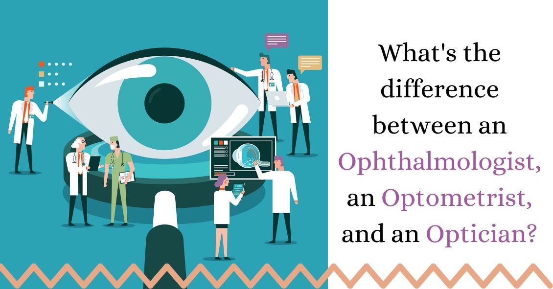
Knowing the difference between the various specialties in the eye care industry can be confusing, especially given the fact that they all start with the same letter and in many ways sound alike.
So, here’s a breakdown of the different monikers to make life a little less confusing for those wanting to get an eye exam.
Ophthalmologists
Ophthalmologists (pronounced “OFF-thal-mologists”) are eye doctors who went to four years of undergraduate university, four years of medical school and four to five years of ophthalmic residency training in the medical and surgical treatment of eye disease.
Many ophthalmologists then go on to pursue sub-specialty fellowships that can be an additional one to three years of education in areas such as cataract and refractive surgery, cornea and external disease, retina, oculoplastic surgery, pediatrics, and neuro-ophthalmology.
Ophthalmologists are licensed to perform eye surgery, treat eye diseases with eye drops or oral medications, and prescribe glasses and contact lenses.
Optometrists
Optometrists are eye doctors who went to undergraduate university for four years, then went on to optometry school for four years.
Many optometrists choose to pursue an additional year of residency after optometry school, though this is not a requirement for licensure. Optometrists are licensed in the medical treatment and management of eye disease, and prescribing glasses and contact lenses.
In some states, optometrists can perform certain minimally invasive laser surgical procedures, but on the whole, optometrists do not perform eye surgery. In addition, optometrists usually have different sub-specialties than ophthalmology, including vision therapy, specialty contact lenses, and low vision.
The analogy I use most often in comparing optometrists to ophthalmologists is that of a dentist and oral surgeon. Many people choose to have optometrists as their primary eye care provider doctor for medical treatment of eye disease, but when surgery is needed, they are referred to the proper ophthalmologist.
Opticians
Opticians specialize in the fitting, adjustment, and measuring of eye glasses. Some states require that opticians are licensed, and others do not.
If you have any questions about which professional is the right fit for your needs, check with your eye-care professional’s office and they’ll be happy to answer them for you.
Article contributed by Dr. Jonathan Gerard
This blog provides general information and discussion about eye health and related subjects. The words and other content provided on this blog, and in any linked materials, are not intended and should not be construed as medical advice. If the reader or any other person has a medical concern, he or she should consult with an appropriately licensed physician. The content of this blog cannot be reproduced or duplicated without the express written consent of Eye IQ.
Read more: Differing Functions of Ophthalmologists, Optometrists, and Opticians

Not everyone understands the importance of sunglasses when the weather turns cold.
Polarized sunglasses are usually associated with Summer, but in some ways it is even more important to wear protective glasses during the Winter.
It’s getting to be that time of year when the sun sits at a much different angle, and its rays impact our eyes and skin at a lower position. This translates to an increase in the exposure of harmful UV rays, especially if we are not wearing the proper sunglasses as protection.
Polarized sunglasses, which are much different than the older dye-tinted lenses, are both anti-reflective and UV resistant. A good-quality polarized sunglass lens will protect you from the entire UV spectrum. This not only preserves your vision, but it also protects the skin around the eyes, which is thought to have a much higher rate of susceptibility to skin cancer.
Snow poses another issue that can be countered by polarized sunglasses.
Snow on the ground tends to act as a mirror because of its white reflective surface and this reflection can become a hindrance while driving. The anti-reflective surface of polarized sunglasses helps reduce the glare and gives drivers improved visibility.
Polarized sunglasses come in many different options based on a patient’s needs. Whether it’s single-vision distance lenses, bifocals, or progressive lenses, there is a polarized lens for every patient.
Winter is a great time of year to ask your optical department about purchasing polarized sunglasses.
Article contributed by Richard Striffolino Jr.
The content of this blog cannot be reproduced or duplicated without the express written consent of Eye IQ

